Cell Counting Application
Our denoising technology enhances fluorescence microscopy images for accurate cell counting and analysis.
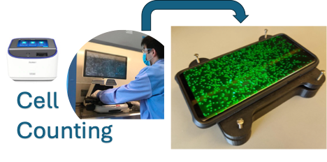
Fluorescence Microscopy Cell Counting
Our technology enhances the quality of fluorescence microscopy images, enabling more accurate cell counting and analysis for research and diagnostic applications.
How It Works
Our advanced algorithm combines histogram thresholding and Deep Image Prior techniques to remove noise from microscopy images.
Image Upload
Upload your fluorescence microscopy images in various formats including JPG, PNG, and TIFF.
AI Processing
Our algorithm automatically processes your image using advanced denoising techniques based on Deep Image Prior networks.
Interactive Comparison
Compare the original and processed images side by side with our interactive slider to see the improvements in detail.
Analysis
Get detailed information about your images including dimensions, noise levels, and quality metrics to understand the improvements.
Our Technology
Learn about the advanced Deep Image Prior (DIP) techniques we use to enhance your images.
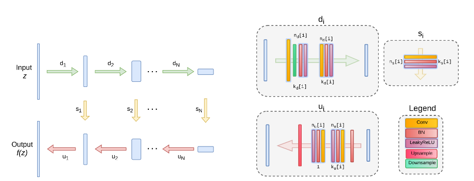
Deep Image Prior Architecture
Our denoising technology leverages Deep Image Prior (DIP), an advanced neural network approach that doesn't require pre-training on large datasets. Instead, it uses the structure of the network itself as a prior for image restoration.
The network architecture shown here includes skip connections, convolutional layers, and specialized components that work together to progressively remove noise while preserving important image details.
Histogram Thresholding
Our system uses advanced histogram thresholding techniques to separate signal from noise in fluorescence microscopy images. This approach is particularly effective for images with distinct intensity distributions between the background noise and the fluorescent signals of interest.
Adaptive Processing
Our algorithm based on DIP and Histogram, adapts to different types of microscopy images and noise patterns. It automatically detects the characteristics of your specific image and applies the optimal combination of processing techniques to achieve the best outcome based on input.